

Introduction:
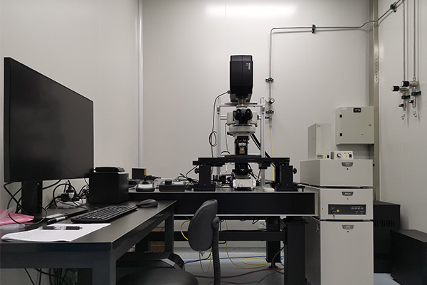
The Nikon A1R manual upright confocal microscope can be used for brightfield, fluorescence and confocal imaging with steady-state imaging resolution XY ≤ 150 nm and Z≤ 300 nm (depending on the emission wavelength). It consists of two sets of electric Z-axis control systems. One of which is independent movable Z-Deck with ±0.2μm repeatability, 10nm step accuracy and 65mm×65mm moving range (XY); the other one is objective lens Piezo fast piezoelectric Z-axis with 1.25nm stepping accuracy, 5nm repeatability and 120Hz frequency, which can perform well in three-dimensional imaging for samples with high time resolution and fast switching speed and accurate position of Z-axis. The electric Z-axis also can be used for large-image stitching, which is convenient for taking the whole morphology of samples. The confocal microscope is equipped with a water objective with high numerical aperture for in vivo imaging of small animals. Also, it is equipped with resonant scanning mirror, which scanning speed of up to 15fps@1024×1024, direction.
Application:
Large field mosaic imaging with high resolution can be performed on stained marker samples in vitro. Living imaging can also performed on the small animal model such as large or small mice, which can reach high spatiotemporal resolution in the awake, behaving animals and living cells. Furthermore, its applications include rapid change detection of blood velocity, ionic concentration and so on. It can achieve precise positioning in any designated area at a single cell level, which is contribute to conducting FRAP on the single cell.
Technical indicators:
Laser:405nm(50mW)、488nm(50mW)、561nm(50mW)、640nm(50mW)
Imaging model: transmitted light brightfield imaging and fluorescence imaging(DAPI、FITC、TxRed、mCherry).
Detector: four channels with assigned detectors.
1ch: DAPI PMT; 2ch: FITC GaAsP PMT;
3ch: TxRed GaAsP PMT; 4ch: mCherry PMT。
Objectives (new arrival):
CFI Plan Apo 10× (Air, NA=0.45, WD=4.0 mm);
CFI Plan Apo 20× (Air, NA=0.75, WD=1.0 mm);
CFI Plan Apo 40× (Air, NA=0.95, WD=0.25-0.16 mm, the thickness of cover glass can be corrected from 0.11 to 0.23 mm);
CFI Plan Fluor 40× (Oil, NA=1.3, WD=0.24 mm);
CFI75 Apo 25× CW 1300 (Water, NA=1.10, WD=2.00 mm), used for confocal and multiphoton;
CFI: Chromatic Aberration Free Infinity
Resolution: the resolution of XY≤150nm, the resolution of Z ≤300 nm (depending on the λ of emission).
Scan resolution: The highest scan resolution can reach 4096×4096; the continuous range of scan zoom is 1~1000×.
Scan mode: Galvano scan mode: 10fps@512×512;Resonant scan mode: 30 fps@512×512.
Image processing software: NIS-Elements C-ER Processing software, including functions of resolution improvement with 1.5 times increase of XY axis and 1.7 times increase of Z axis,3D visualization, intracellular structure measurement,fluorescence intensity measurement,colocalization analysis.


Address :Chinese Institute for Brain Research, Beijing(CIBR), Jianzan Building, No.26, Science Park Road, Zhongguancun Life Science Park, Changping District, Beijing, China
-
Contact:010-80765986 Postalcode:102206
© 2023 by Personal Life Coach. Proudly created with Wix.com ICP备案号:京ICP备18029179号