

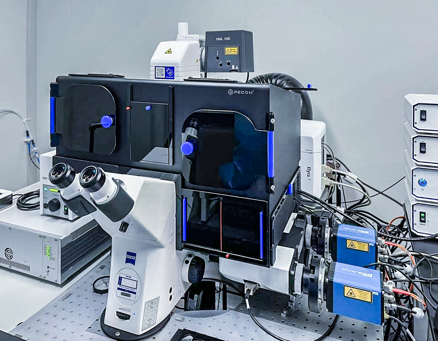
Zeiss Inverted Structured illumination Microscope (Elyra 7)
Room: X219->X214
Introduction
Structured Illumination Microscopy (SIM) improves resolution by utilizing non-uniform excitation light patterns with sinusoidal intensity variations and powerful image reconstruction techniques. The Elyra 7 uses Lattice SIM technology, which employs lattice patterns (rather than grid lines) to illuminate the sample area. Compared to traditional SIM, Lattice SIM has the advantage of fast imaging speed and high photon utilization, making it suitable for imaging living cells and obtaining high-resolution images of conventional thin samples. Elyra 7 is powered by a new, groundbreaking two-step image reconstruction algorithm called SIM². In the first step, diffraction order combination, noise removal and frequency suppression filtering are applied. These digital image operations produce a digital SIM point spread function (PSF). Subsequent iterations of deconvolution use this PSF. Similar to using experimental PSF for hardware-based microscopic data deconvolution, the SIM² algorithm outperforms traditional single-step image reconstruction methods in terms of resolution, optical cross-section, and stability. SIM² improves the resolution of structured light illumination microscope data and the quality of the optical section. Elyra 7 offers a SIM Apotome mode that uses grid lighting and swiftly adjusts fluorescence signals in various focal planes. By capturing three or five images of different grid positions (phases), the imaging software combines these frames into a single image that contains only the focal plane information (optical section image). This provides a fast optical section image with high contrast and resolution (both transverse and axial). Additionally, Elyra 7 offers Burst mode and Leap mode to further improve imaging speed. Burst mode processing uses a rolling window method that enables you to view live sample processes at up to 255 fps. Leap mode only requires imaging every three planes, reducing imaging time and reducing photobleaching of the sample. Elyra 7 is equipped with a Single-Molecule Localization Microscopy (SMLM) module. In SMLM, fluorescent molecules are randomly activated so that only one of the many molecules is active. This enables the determination of its center of mass with positioning accuracy above the diffraction limit of the point diffusion function. Once recorded, the molecules will go dark, and the activation and shutdown process will be repeated until all the molecules are captured. The positioning information can then be plotted in a new image to create an ultra-high resolution image. Elyra 7's SMLM technology, such as PALM, dSTORM, and PAINT, can achieve 20-30 nm landscape resolution.
Application:
The Elyra 7 inverted microscope is versatile and compatible with a range of scenes, from wide field to super resolution, for both live cells and conventional sections. It supports common dyes and fluorescent proteins and offers the advantages of fast shooting speed and low light damage. The live cell incubation system is designed to accommodate 35mm and 60mm petri dishes as well as standard slides. Moreover, the O2 control module can create a suitable growth environment for anaerobic cells.
Technical indicators:
Laser:405nm(50mW), 488(100mW), 561nm(100mW),
642nm(500mW).
Objectives:
10X (Air, NA=0.30, working distance 5.20mm)
10X (Air, NA=0.45, working distance 2.00mm)
20X (Air, NA=0.8, working distance 0.55mm)
25X (Water, oil, glycerin, silicone oil, NA=0.8, working distance 0.57mm)
40X (Water, NA=1.2, working distance 0.28mm)
63X (Oil, NA=1.4, working distance 0.19mm)
63X (Oil, NA=1.46, working distance 0.10mm)
Imaging model: Lattice SIM, SIM Apotome, SMLM and TIRF.
Filter Parameters: DAPI: EX 365, BS FT 395, EM BP 445/50
GFP/mRFP/Alexa 633: EX TBP 483+564+642, BS TFT 506+582+659, EM TBP 526+601+688
Detector: sCMOS high-speed imaging dual camera,
physical pixel 2048×2048, pixel size 6.5µm×6.5µm. QE 82%, sampling depth 15bit.
Scan magnification: 1x and 1.6x
Scan resolution: 4×1~8192×8192. The scan resolution of all channels can reach 8192×8192 and 16 bits grayscale depth.
Live cell incubation system:
Temperature control range: room temperature to 60℃, temperature adjustment accuracy is 0.01℃;
CO2 control range: automatic mixing concentration control range 0 to 8%, adjustment accuracy 0.01%;
O2 control module: automatic mixing concentration control range 0 to 21%, adjustment accuracy 0.01%;
Humidity control range: 50%-70% (37℃).
Adapter: It can be matched with 35mm and 60mm petri dishes, standard slides, small eight-hole culture tanks, porous plates.
Image processing Software:
Zeiss ZEN - can assist you in completing the entire process from image acquisition to data analysis, including but not limited to SIM, SIM2, Burst, SMLM algorithm processing.
Arivis - Visualizes and quantifies large 3D and 4D datasets derived from imaging. Arivis Vision4D not only renders volumetric images of virtually unlimited size but also provides advanced image processing tools such as volume fusion, channel offset correction, traditional and machine learning-based segmentation, and 3D tracking. You can visualize your quantitative results in the Arivis Vision4D software or export all data for further analysis. The Arivis Vision4D module structure can be flexibly adapted to your needs for advanced image processing and analysis.

Address :Chinese Institute for Brain Research, Beijing(CIBR), Jianzan Building, No.26, Science Park Road, Zhongguancun Life Science Park, Changping District, Beijing, China
-
Contact:010-80765986 Postalcode:102206
© 2023 by Personal Life Coach. Proudly created with Wix.com ICP备案号:京ICP备18029179号