

Instrument Name:
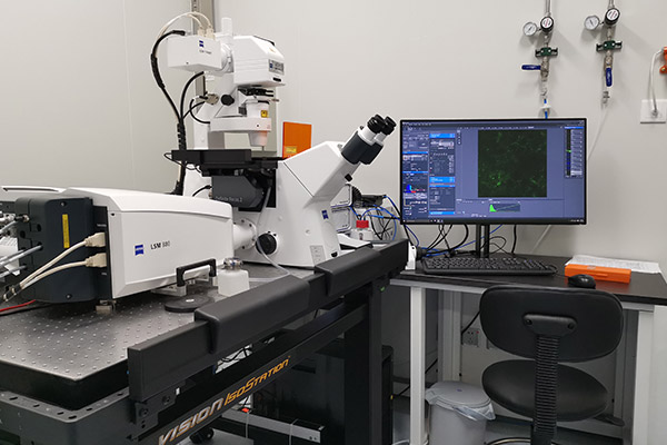
Zeiss Inverted Confocal Microscope (LSM 880) Room: X208
Introduction
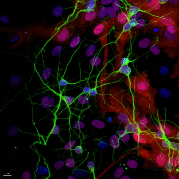
The Zeiss LSM 880 inverted confocal fluorescence microscope is mainly used for long-term bright-field and fluorescence imaging of cultured cells or tissues. The system is equipped with Zeiss's unique Airyscan detector, which improves both resolution and imaging speed. Airyscan has a set of 32-array GaAsP-PMT detectors without traditional pinholes; the 32 micro-units act like 32 pinholes, each sensing light from the sample, and an algorithm re-sums and re-positions the azimuthal signals to form a correct, clear image. Without a pinhole, more light is collected—1.25× that of a traditional pinhole (1.25 AU), each micro-unit equivalent to 0.2 AU—greatly improving SNR and enhancing resolution and detail brightness. At 488 nm, Airyscan's detection capability is 1.7× that of traditional confocal, with XY resolution up to 140 nm and Z resolution up to 400 nm.
Application
The inverted microscope is suitable for observing living cells in culture dishes, equipped with DIC imaging mode for transparent cells. The live-cell incubation system accommodates 35 mm and 60 mm dishes and standard slides, enabling long-term continuous imaging of live cells and high-resolution imaging of fixed samples. It is suitable for observing and recording brain tissue sections, fluorescent labeling of live cells, 3D reconstruction, and qualitative, quantitative, timed and localized distribution detection of cellular substances and ions, thereby analyzing physiological and biochemical changes in brain tissue, cells or between cells.
Key Technical Specifications
Laser:
405nm(30mW), 561nm(20mW), 633nm(5mW), multiline argon-ion green laser 458/488/514nm(25mW); fast AOTF, 8 channels, switching time 5 µs
Objectives:
10x (Air, NA=0.45); 20x (Air, NA=0.8); 25x (Water/Glycerin/Silicone/Oil, NA=0.8, WD=0.57mm); 40x (Air, NA=0.6, WD=3.48mm); 40x (Oil, NA=1.3, WD=0.2mm); 63x (Oil, NA=1.4, WD=0.19mm)
Imaging Mode:
Transmitted brightfield, DIC, and fluorescence imaging (DAPI, GFP, Cy3, Cy5); minimum spectral detection interval 3 nm
Filter Sets:
DAPI: EX 365, BS FT 395, EM BP 445/50; GFP: EX BP 470/40, BS FT 495, EM BP 525/50; CY3: EX BP 545/25, BS FT 570, EM BP 605/70; CY5: EX BP 640/30, BS FT 660, EM BP 690/50
Detectors:
Two built-in GaAsP ultra-high-sensitivity linear detectors, two PMT linear detectors, one high-sensitivity monochrome cooled CCD; one GaAsP serves as the Airyscan detector
Resolution:
Super-resolution mode XY 120 nm, Z 350 nm, 5 fps (512×512, 16-bit); fast SR mode X 145 nm, Y 180 nm, Z 450 nm, 19 fps (512×512, 16-bit)
Scan Resolution:
Freely selectable from 4×1 to 8192×8192; each channel reaches 8192×8192 with 16-bit grayscale
Scan Speed:
Galvanometer: 13 fps (512×512, 16-bit), 430 fps (512×16, 16-bit); line scan: 4–6875 lines/s (512×1, 16-bit); fast scan: 27 fps (488×488, 16-bit); CCD frame: 38 fps (1936×1460)
Spectral Range:
400 nm ~ 1000 nm; sampling depth 14-bit
Scan Zoom:
Minimum optical zoom 0.6x ~ 40x
Live Cell Incubation:
Temperature control room temperature to 60 ℃, accuracy 0.01 ℃; CO2 auto-mixing 0–8%, accuracy 0.01%
Humidification:
200 ml pure-water large-capacity humidifier (heatable); incubation chamber fits 35/60 mm dishes, standard slides, 8-well chambers
Image Processing Software:
Spectral scanning and unmixing (removes autofluorescence/crosstalk), free large-image stitching, co-localization and basic measurement; physiology module (ROI mean fluorescence, real-time/post-scan ion concentration), histogram tools (length/angle/area/intensity)


Address :Chinese Institute for Brain Research, Beijing(CIBR), Jianzan Building, No.26, Science Park Road, Zhongguancun Life Science Park, Changping District, Beijing, China
-
Contact:010-80765986 Postalcode:102206
© 2023 by Personal Life Coach. Proudly created with Wix.com ICP备案号:京ICP备18029179号