

Instrument Name:
Olympus VS120 Virtual Slide Microscope (VS120-S6-W) Room: 1024
Introduction
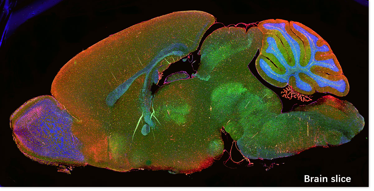
The VS120 not only creates high resolution brightfield images, but also can scan in full multi-fluorescence mode (4 channels). Scanning multiple large specimens up to 31 Z-planes enables it to be a powerful tool for viewing thicker specimens, such as cell clusters or brain slices. An innovative algorithm makes image stitching more precise, enabling high-level accuracy from small animal brain slices to large specimens. Thus, the VS120 switches seamlessly between micro and macro observation to enable swift viewing of regions of interest and overall structures.
Application
This system is mainly used to scan specimens on glass slides, such as brain slices and other tissue slices. The efficiency of scanning is greatly improved by the 6-slide motorized stage. It is also equipped with a 2″×3″ stage suitable for observing larger specimens.
Key Technical Specifications
Fluorescence Channels:
Cy5: Ex. 628/40 nm; Em. 692/40 nm Texas Red: Ex. 586/20 nm; Em. 628/32 nm Cy3: Ex.540/25 nm; Em.605/55 nm (new) GFP: Ex. 472/30 nm; Em. 520/35 nm DAPI: Ex. 365/10 nm; Em. 440/40 nm
Observable Specimen:
Glass slide with cover glass (26 mm x 76 mm)
Scan Area:
Single slide 26 mm x 64 mm
Scan Time:
Approx. 2 min. (20x objective, scan area 15 mm x 15 mm brightfield)
Scan Resolution:
10x (NA 0.4): 0.66 μm/pixel; 20x (NA 0.75): 0.33 μm/pixel; 40x (NA 0.95): 0.17 μm/pixel
Objectives:
2x, 10x, 20x, 40x
Cover Glass Thickness:
0.12 mm – 0.17 mm
Stage:
6-slide motorized stage, two 2″×3″ slides stage


Address :Chinese Institute for Brain Research, Beijing(CIBR), Jianzan Building, No.26, Science Park Road, Zhongguancun Life Science Park, Changping District, Beijing, China
-
Contact:010-80765986 Postalcode:102206
© 2023 by Personal Life Coach. Proudly created with Wix.com ICP备案号:京ICP备18029179号