

Instrument Name:
Opera Phenix High-throughput and High-content Imaging Analysis System Room: X213
Introduction
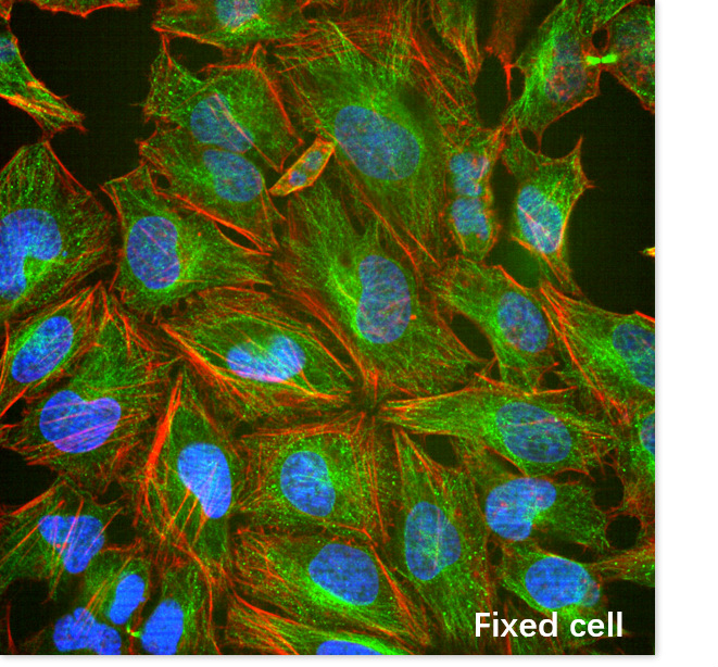
The Opera Phenix high-throughput and high-content imaging analysis screening system is a new generation of imaging system based on Nipkow dual-disk scanning laser confocal. The dual-disk structure supports synchronous multi-color confocal image acquisition, minimizes spectral cross-interference, improves imaging speed and sensitivity, and reduces the impact of phototoxicity and photobleaching on live-cell observation while improving light efficiency. The system is equipped with a water objective and a fully automatic objective water-replenishment circulation system; the water objective has high light efficiency and requires shorter exposure time, making it more suitable for long-term live-cell detection and high imaging quality in thick samples. The DPC imaging mode obtains richer cell biology information.
Application
The system provides a variety of solutions for automatic, high-speed and high-resolution imaging screening, meeting the needs of drug discovery and high-throughput biology. It is suitable for cell screening at subcellular resolution and various bead-based screening applications ranging from conventional microplates to micro-nano plates. Different analysis methods—fluorescence intensity regional analysis, cell morphology analysis, intracellular texture (cytoskeleton, organelle mitochondrial distribution, etc.), and label-free bright-field analysis—are integrated to comprehensively analyze cell state, changes and overall trends, yielding high-content, high-reliability results that characterize cells from multiple dimensions. It is widely applied in apoptosis, cell cycle, cytotoxicity, receptor translocation, protein interaction, and is an important tool in cell biology, systems biology, cancer research, cardiovascular research, stem cell research and neuroscience.
Key Technical Specifications
Light Source:
Built-in 4-color solid-state lasers: 405nm, 488nm, 561nm, 640nm; near-infrared LED as bright-field light source
Imaging Modes:
Wide-field fluorescence, confocal, bright-field, and label-free digital fluorescence (DPC) imaging
Channels:
DAPI: EX 405nm, EM 435-480nm; GFP: EX 488nm, EM 500-550nm; CY3: EX 561nm, EM 570-630nm; CY5: EX 640nm, EM 650-760nm
Detector:
Built-in high-sensitivity high-resolution synchronous detector (dual sCMOS cameras, 16-bit), simultaneously acquiring fluorescence and bright-field images at different wavelengths; daily imaging throughput ≥ 100,000 frames
Objectives:
1.25x (Air, NA=0.03, WD=4); 5x (Air, NA=0.16, WD=12.1); 10x (Air, NA=0.3, WD=5.2); 20x (Air, NA=0.4, WD=8.28, Correction collar); 20x (Air, NA=0.8, WD=0.55); 20x (Water, NA=0.8, WD=0.55); 40x (Air, NA=0.6, WD=3.28, Correction collar); 40x (Water, NA=1.1, WD=0.62); 63x (Water, NA=1.15, WD=0.6)
Motorized Scanning Stage:
High-precision magnetic-suspension encoded electric XY stage (step accuracy 50 nm, repeatability 100 nm)
Adapters:
SBS standard microplates of 6, 24, 48, 96, 384 and 1536 wells, plus user-defined plates and slides; slide adapter (4 pieces)
Environmental Control Unit:
Temperature control 37-42 (±1) °C; CO2 concentration adjustable 1-10% (±0.5%)
Image Processing Software:
Harmony acquisition and full-function module analysis software, including 25 analysis modules and 37 preset application solutions; texture analysis (8 texture filters), machine self-learning, STAR analysis (207 morphological parameters), data clustering analysis, label-free DPC module, and 3D cell observation module with built-in 3D co-localization analysis


Address :Chinese Institute for Brain Research, Beijing(CIBR), Jianzan Building, No.26, Science Park Road, Zhongguancun Life Science Park, Changping District, Beijing, China
-
Contact:010-80765986 Postalcode:102206
© 2023 by Personal Life Coach. Proudly created with Wix.com ICP备案号:京ICP备18029179号