

Instrument Name:
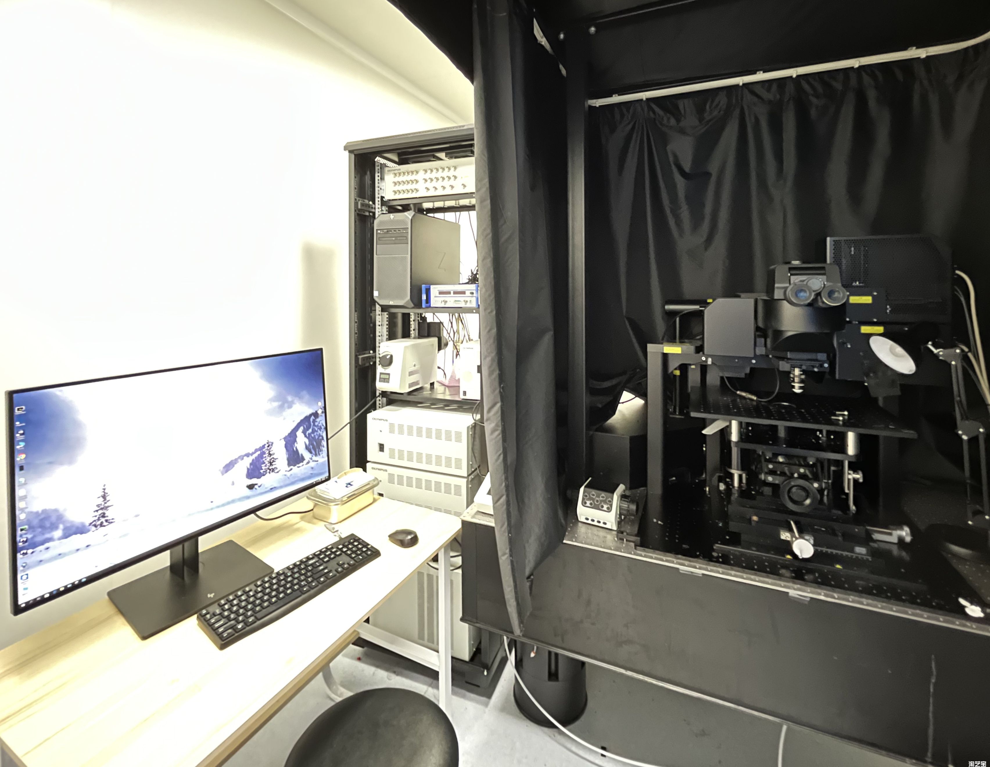
Olympus FVMPE-RS Single-Line Two-Photon Upright Microscope Room: B311
Introduction
The FVMPE-RS combines laser scanning confocal microscopy with two-photon excitation technology, enabling long-term imaging of live samples. Its unique four-axis automatic correction automatically adjusts the laser angle and position when any laser wavelength is changed, so that the two spots of multi-color lasers completely coincide. It is equipped with analog and digital signal converters for electrophysiology; the ANALOG converter synchronizes external devices such as electrophysiology equipment with two-photon imaging, laser stimulation, and other functions. External experimental devices (e.g. electrophysiology equipment) can be triggered through input or output signals, supporting TTL and I/O signals. The Analog unit can also convert voltage into images via software, and can do the same for fluorescence images, synchronizing the electrical signal measured by the patch clamp during light stimulation with the acquired image and displaying it as pseudo-color of light intensity. The image workstation and analysis software can control all hardware devices, including functions such as Analog external trigger signal control. For post-acquisition results, it can perform three-dimensional reconstruction of massive data, 3D surface rendering, zooming, rotation display, and other image-processing functions. Using femtosecond lasers as the light source, it can penetrate deep into tissues and nonlinearly excite fluorescent molecules; the long wavelength reduces laser-induced damage to organisms and lowers phototoxicity. The two-photon system features a Deep Focus Mode that achieves deeper imaging than the traditional mode. While ensuring high spatial resolution (imaging depth can exceed 1 mm), the instrument also supports long-term live imaging.
Application
The system features high imaging depth and low phototoxicity, making it suitable for long-term real-time imaging of live animals. Because two-photon excitation stimulates only the focal-plane signal, it is more suitable for precise light stimulation experiments such as light activation and photobleaching.
Key Technical Specifications
Laser:
Single line IR laser: 800-1300 nm wavelength tunable
Objectives:
5x (Air, NA=0.1, WD=20 mm); 10x (multi-immersion adjustable, NA=0.6, correction collar (cover glass: 0-0.23 mm, n: 1.33-1.52), WD=8 mm); 25x (Water, NA=1.05, correction collar (cover glass: 0-0.23 mm), WD=2 mm)
Imaging modes:
widefield fluorescence imaging, brightfield imaging, two-photon imaging
Stage:
manual stage, adjustment range: 125-195 mm
Fast piezo Z-axis:
Z-axis travel range: 400 µm; Z-axis stepping accuracy: 1.25 nm; repeatability: 5 nm; frequency: 120 Hz
Detector:
4-channel fixed detector: external detector (two-photon): 2 GaAsP (ultrasensitive detector), 2 PMT (photomultiplier tube)
Scan mode:
high-resolution (Galvano-mirror) and high-speed scanning mode (Resonant-scanner) selectable; in high-speed mode, scanning speed 30 fps @ 512×512, 438 fps @ 512×32
Analog and digital signal converters for electrophysiology:
6 TTL signal inputs, 6 TTL signal outputs, 4-channel analog signal input port, 1-channel analog signal output port
Image processing software:
FVMPE-RS basic software


Address :Chinese Institute for Brain Research, Beijing(CIBR), Jianzan Building, No.26, Science Park Road, Zhongguancun Life Science Park, Changping District, Beijing, China
-
Contact:010-80765986 Postalcode:102206
© 2023 by Personal Life Coach. Proudly created with Wix.com ICP备案号:京ICP备18029179号