

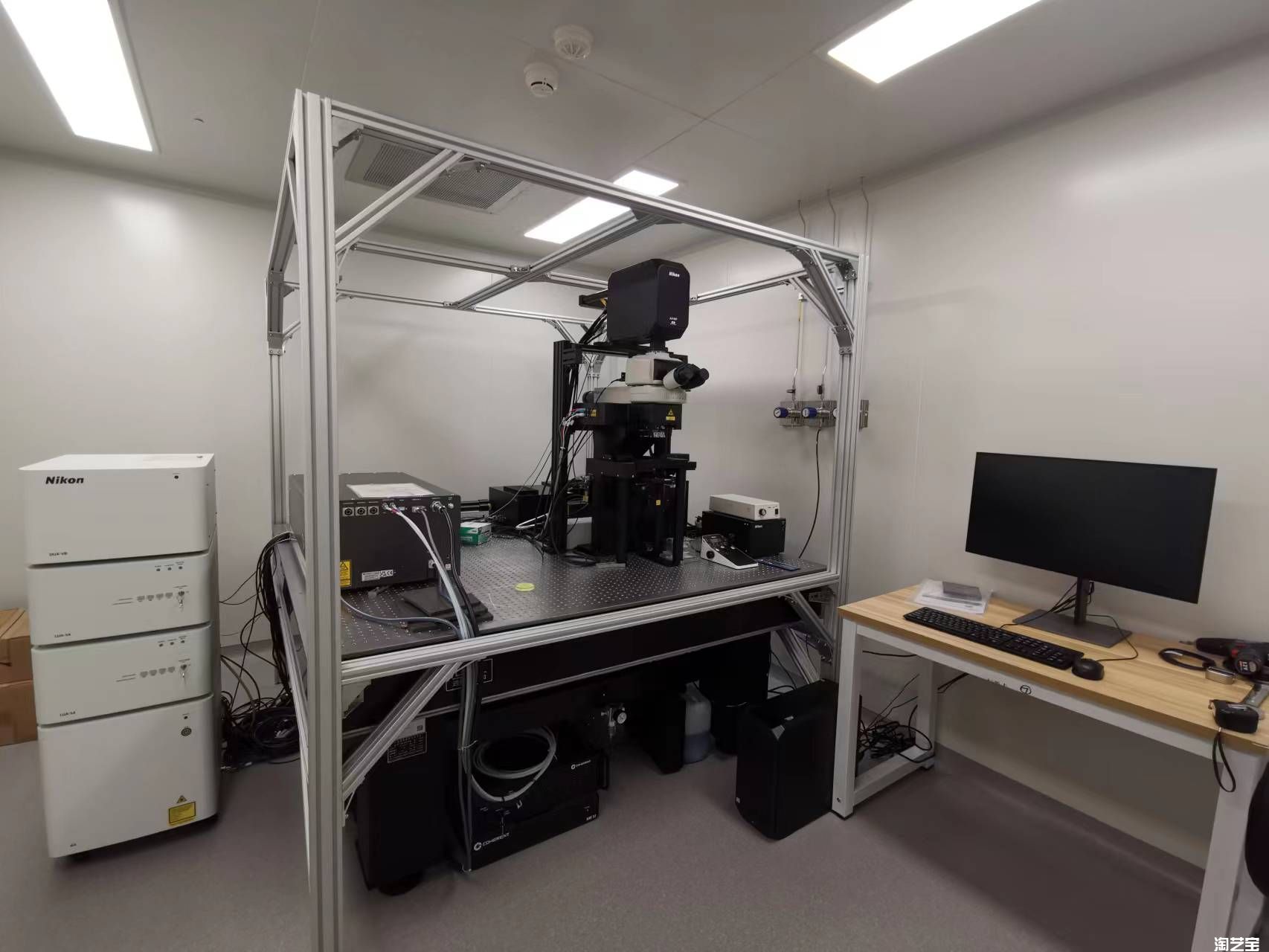
Nikon Two-photon upright microscope (AX R MP)
Room:X219>X216
Introduction
Combining the technology of laser scanning confocal microscopy and two-photon excitation technology, AX R MP can penetrate deep inside the tissue and excitation of fluorescent molecules. The long wavelength can reduce the damage of the laser on the organism, reduce phototoxicity, as well as ensure high spatial resolution (imaging depth can exceed 1mm) at the same time. The device can also be used alone with single photon imaging capabilities.
Utilizing Nikon's new CFI75 tilting nosepiece, the objective can be oriented to image samples in their natural positions, minimizing stress and uncontrolled variables. This nosepiece is compatible with Piezo Z, maximizing 3D image speed in these challenging samples. Both galvanometer mode and resonance mode can obtain a field of view of 22mm diagonally, and the acquisition speed is faster than that of the previous generation A1. The AI de-noise function can obtain images with high signal-to-noise ratio while fast acquisition. The fast piezo Z axis can be used for 3D imaging of samples requiring high time resolution and fast and accurate switching of Z axis. The space below the microscope objective is 40cm high, providing sufficient free space for in vivo observation, which not only improves the flexibility of sample placement, but also facilitates user operation.
Application:
The system has the characteristics of high imaging depth and low phototoxicity, which is suitable for long-term real-time imaging of live animals. Two photons can only stimulate the focal plane signal principle characteristics, can be more accurate light activation, photo bleaching and other light stimulation experiments. The tunable wavelength laser can be used for multi-standard sample imaging. It is equipped with two sets of single photon lasers, which support synchronous optical stimulation and image acquisition in the optical stimulation experiment. AX R MP is equipped with high numerical aperture water lens which is suitable for in vivo imaging of animals.
Technical indicators:
Laser: Continuous Wave Laser (2 sets): 405nm, 488nm, 561nm,
640nm; Femtosecond pulse laser: 1040nm, adjustable wavelength
680-1300nm
Imaging model: Wide-field fluorescence imaging, bright-field imaging,
single-photon confocal imaging, two-photon imaging
Detector: 4 highly sensitive detectors GaAsP; External detectors: 4 NDD (ultra-sensitive detector), 2 spectral scanning PMTS (photomultiplier tube)
Objectives: CFI Plan Apo 10× (Glyc, NA=0.5, WD=5.5, 400-1300 nm, clarity);
CFI Plan Fluor 10× (Water, NA=0.3, WD=3.5, 400-1000 nm);
CFI Apo 20× (Water, NA=1.0, WD=2.8, 400-1300 nm);
CFI Apo 25× (Water, NA=1.1, WD=2.0, 400-1300 nm);
CFI Apo NIR 40× (Water, NA=0.8, WD=3.5, 400-1000 nm);
CFI Apo NIR 60× (Water, NA=1.0, WD=2.8, 400-1000 nm);
CFI Plan 100× (Water, NA=1.1, WD=2.5, 400-1000 nm);
CFI Plan Apo 25× (Silicone oil, NA=1.05, WD=0.55, 400-1000 nm);
CFI Plan Apo 40× (Silicone oil, NA=1.25, WD=0.30, 400-1000 nm);
CFI Plan Apo 2 x (Air, NA=0.1, WD=8.5, 400-1000 nm);
CFI Plan Apo 4× (Air, NA=0.2, WD=20, 400-1000 nm);
CFI Plan Apo 10× (Air, NA=0.45, WD=4.0, 400-1000 nm);
CFI Plan Apo 20× (Air, NA=0.8, WD=0.8, 400-1000 nm);
CFI Plan Apo 60× (Oil, NA=1.42, WD=0.15, 400-1000 nm);
CFI Plan Apo 100× (Oil, NA=1.45, WD=0.13, 400-1000 nm);
Resolution: In confocal mode, imaging resolution XY≤150nm and Z-axis ≤300 nm (depending on emission wavelength).
Scan resolution:The maximum scanning resolution should not be less
than 8192×8192 image decimal point; Optical
doubling: ≥1 ~ 1000X (continuous change).
Scan mode:Galvo mode :512×512 resolution (4 fluorescent channels
plus 1 transmission channel), speed 2 fps; Resonate Mode: 2048 x 16 resolution,
720 fps.
Scan zoom: ≥1 ~ 1000X (continuous change).
Mobile range of electric loading platform: ±34 mm (X) and ±27 mm (Y)
Image processing software: NIS-Elements C-ER Processing software, including
functions of resolution improvement with 1.5 times increase of XY axis and 1.7
times increase of Z axis,3D visualization,
intracellular structure measurement,fluorescence intensity
measurement, colocalization analysis.

Address :Chinese Institute for Brain Research, Beijing(CIBR), Jianzan Building, No.26, Science Park Road, Zhongguancun Life Science Park, Changping District, Beijing, China
-
Contact:010-80765986 Postalcode:102206
© 2023 by Personal Life Coach. Proudly created with Wix.com ICP备案号:京ICP备18029179号